PRODUCT DESCRIPTION
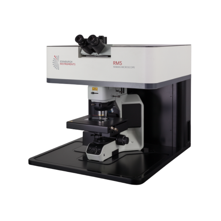
The RM5 is a compact powerhouse designed for every laboratory, from industry to academia. With advanced spectral imaging capabilities, intuitive operation, and precise confocal optics, the RM5 delivers unparalleled performance across diverse applications such as materials science, pharmaceuticals, and beyond.
Featuring a simple yet comprehensive design, the RM5 ensures ease of operation for users at any level of experience with Raman microscopy. Ramacle® software guides users through all measurement stages without limiting any Raman capabilities, resulting in the creation of high-quality, publication-ready data.
Configurable with up to three fully software-controlled internal lasers spanning 405 nm – 1064 nm, this system maximises experimental range while maintaining a compact footprint. Laser selection is complimented by a 5 position grating turret, where gratings are selected to perfectly match chosen lasers. This design not only saves laboratory space but also enhances ease of use, eliminating the need for users to manually activate lasers or exchange gratings.
The RM5 enhances user experience with key features: it includes an internal silicon standard for automatic wavelength calibration, ensuring precise spectral measurements without user intervention. The confocal microscope offers the most pinhole positions available, providing flexibility for optimal measurement conditions. When integrated with a motorised stage, the software generates both 2D and 3D Raman and photoluminescence maps, along with SurfMAP® for mapping uneven samples.
This versatility allows the RM5 to seamlessly integrate into any laboratory setting, accommodating a wide range of experiments across disciplines from biologists to chemists to physicists, making Raman microscopy accessible to all.
SOFTWARE
Ramacle® is an all-encompassing software package written for ease of use and complete operation of the RM5.
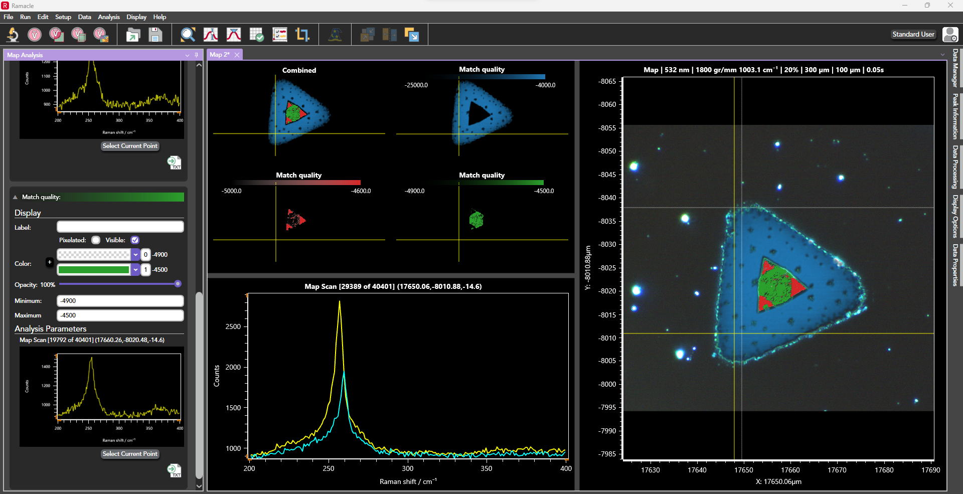
The software prioritises automation, reducing the need for manual adjustments and allowing users to focus more on sample analysis. From initial microscope setup, where users visualise their sample in a spacious window, to configuring laser parameters, pinhole settings, and grating selection for spectral acquisition, Ramacle handles all aspects seamlessly.
On the RM5, Ramacle comes with standard data acquisition methods such as single measurements, multiple and accumulated scans, kinetic scan, as well as a comprehensive suite of mapping techniques when upgraded to a motorised stage e.g. 2D, 3D, SurfMAP, and FastMAP. Upgrades and accessories such as temperature stages and multiwell plates unlock additional software features.
Spectral Measurements
Intuitive use is at the core of Ramacle’s design. We begin by focusing your sample using the microscope setup. Once you’re satisfied with the microscope image, we can proceed to the measurement setup window. The white-light image will carry over, allowing you to click on different areas of the sample for live Raman feedback using the live option. This feature helps you verify your measurement parameters and focus. You can then take single or multiple spectra from the selected sample area.

Figure 1: Ramacle Software
Additionally, kinetic series can be set up simply in Ramacle, and if using a temperature stage, temperature ramps and spectral series can also be set. The RM5 can be configured to acquire photoluminescence emission spectra. Ramacle measures spectral PL responses using the CCD camera enabling single point and mapping data to be collected. Measurements are made by selecting a low groove density grating to cover the entire emission range. The software can be operated in both wavenumber and wavelength scale making working in Raman and PL effortless.
3D Mapping
Ramacle operates 3D mapping similarly to 2D mapping, with the added ability to define the map along the Z-axis. Thanks to the truly confocal nature of the RM5, highly resolved 3D maps can be captured. This advanced capability extends Raman mapping beyond surface analysis, constructing a detailed 3D chemical image at the micron scale.
The Ramacle software can then display the result focusing on one 2D layer, on the 2D stacks collected, or as a full volumetric. Shown below are two common examples for 3D mapping; first 3 µm polystyrene beads, and on the right we a see an inclusion in pegmatite. Green is quartz, red is anatase, and blue is an unknown inclusion.
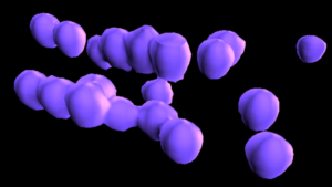
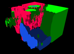
Figure 1: 3D Mapping of polystyrene bead and pegmatite
TECHNICAL SPECIFICATIONS
| LASER SPECIFICATION | |
|---|---|
| Lasers | Up to 3 narrow-band lasers including: 532 nm, 638 nm, 785 nm Other wavelengths available on request Laser selection is fully computer-controlled |
| Laser Rejection Filters | Up to 3 laser rejection filters included Filter exchange is fully computer-controlled |
| Laser Attenuation | 4 orders of magnitude, continuous Fully computer-controlled |
| SPECTROGRAPH | ||
|---|---|---|
| Spectral Resolution | From <0.3 cm-1 | |
| Spectral Range | <50 cm-1 – 15,000 cm-1* | |
| Spectrograph | Type Focal Length Gratings Slits | Asymmetric Czerny-Turner 225 mm 5-position grating turret, fully computer-controlled Continuously adjustable, fully computer-controlled |
| Confocal Imaging | Adjustable confocal pinhole, fully computer-controlled |
| DETECTORS | ||
|---|---|---|
| Detectors | Standard Detector | High sensitivity ultra low noise CCD 1650 x 200 pixels, TE-cooled -60°C (standard) OR 2000 x 256 pixels, TE-cooled -60°C (enhanced sensitivity and spectral range) |
| Optional Second Detector | EMCCD detector, InGaAs and others available on request Selection of detectors, fully computer-controlled |
| SYSTEM | ||
|---|---|---|
| Raman Polarisation | Optional | Polarisation kit available, fully computer-controlled |
| Internal Calibration | Wavelength calibration standard (Neon) Raman shift standard (Silicon) Sensitivity validation standard (Silicon) Automated laser alignment | |
| Microscope System | Functionality | Full upright microscope with brightfield and darkfield illuminator Polarisation, Differential Interference Contrast (DIC) capability and fluorescence imaging 10x and 100x objective included as standard; up to 5 can be included |
| Optional Objective(s) | Trinocular eyepiece, embedded CMOS video camera, second video camera optional | |
| Sample Viewing | XY manual stage | |
| Sample Stage | XYZ motorised stage (75 mm x 50 mm XY), confocal Raman mapping | |
| Optional | Temperature-controlled sample stages available | |
| Software | Ramacle® | Comprehensive all-in-one, intuitive software package |
| Operating System | Windows® | |
| Functionality | Data acquisition, spectrograph control, graphical display, data processing | |
| Optional | Chemometric, spectral library packages – KnowItAllTM | |
| Laser Safety | Without Laser Enclosure With Laser Enclosure | Class 3B Class 1 |
| Dimensions | W x D x H † Weight † | 600 mm x 800 mm x 600 mm † 63 kg * depending on grating, laser and CCD selection † without laser enclosure |