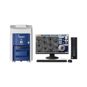
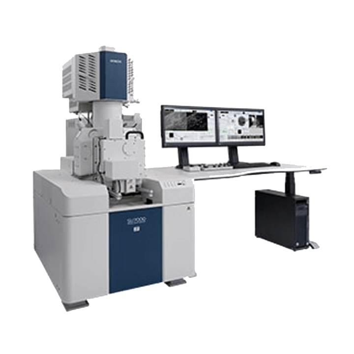
SU7000: The Next-Generation FE-SEM
The modern FE-SEM requires not only high performance but also a multitude of functionalities including wide-area observation, in-situ analysis, variable pressure, high-resolution imaging at low accelerating voltages, and simultaneous multi-signal collection.
The SU7000 is designed to address these aspects and more by delivering enhanced information for diversified needs in the field of electron microscopy.
Experience the nano-world with the SU7000!
Key Concept
1.Versatile Imaging Capability
The SU7000 excels in fast acquisition of multiple signals to address expansive SEM needs, from imaging a wide field of view to visualizing sub-nanometer structures and everything in between.
The incorporation of newly designed electron optics and detection systems allows for efficient simultaneous acquisition of multiple secondary electron and back-scattered electron signals.
2.Multi-Channel Imaging
The number of the detectors mounted on the SEM is ever increasing, along with the need to display all collected information effectively.
The SU7000 is capable of processing, displaying, and saving up to 6 signals simultaneously to maximize information acquisition.
3.Wide Variety of Observation Techniques
The specimen chamber and the vacuum system are optimized for:
- Large specimen size
- Sample manipulation at various axes
- Variable pressure conditions
- Cryogenic conditions
- Heating and cooling in-situ observation
4.Microanalysis
The electron gun is equipped with a Schottky emitter that provides up to 200 nA beam current to accommodate various microanalysis applications.
The specimen chamber and port layout are designed to incorporate multiple analytical options including EDX, WDX, EBSD, cathodoluminescence, and more.
The SU7000 with the combination of numerous analytical accessories unifies multi-discipline techniques in a single platform.
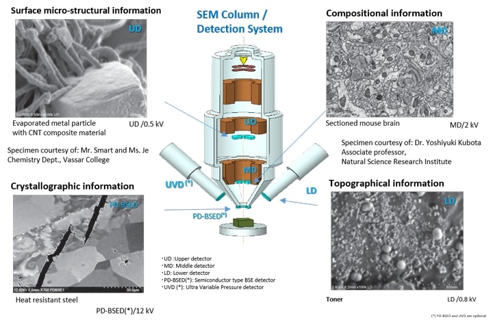
Imaging Performance
Enhanced Information Acquisition
The advanced detection system of the SU7000 streamlines acquisition of structural, topographical, compositional, crystallographic, and other types of information by minimizing changes to microscope conditions, such as working distance or accelerating voltage.
Single-Scan Multi-Signal Imaging
Specimen: Organic-coated gold rods
Specimen courtesy of: Mr. Smart and Ms. Je Chemistry Dept.,
Vassar College
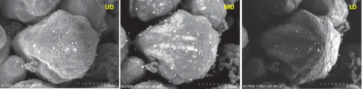
Simultaneous image acquisition for surface micro-structural information (UD), surface coating (MD), and overall topographic information (LD). Acceleration voltage: 1 kV
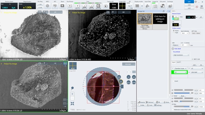
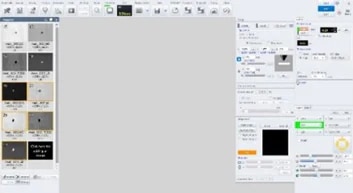
Intuitive Graphical User Interface
Enhanced Signal Display
- Customizable display modes.
- Single and Dual-monitor configurations.
- Simultaneous image display up to 4-ch (single) and 6-ch (dual).
- Chamber Scope and SEM MAP for optical stage navigation.
Highly Flexible Screen Layout
The software is capable of display 1, 2, or 4 signals including the chamber scope or SEM MAP on a single monitor.
Additionally, the operation panel can be customized to display submenus anywhere on the screen.
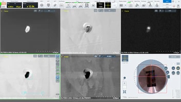
Dual Monitor
The first monitor can be used as a dedicated image display while the second monitor is utilized for operation.
Five detector images (UD, LD, UVD, MD, and PD-BSED) and SEM MAP of non-metallic inclusions in a steel specimen are displayed (left).
The screen shows the operation panel menu and the thumbnail image window on one screen (right).
The dual-monitor configuration supports increased productivity with expanded workspace.
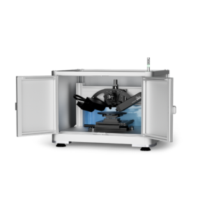
Expandable Observation and Analysis
Large specimen chamber and large stage
The specimen chamber can accommodate a φ 200 mm specimen and 18 accessory ports. The large stage travels 135 mm (X) x 100 mm (Y) and can accept up to 2 kg of specimen.(*) Large specimen or variable type of sub-stages can be easily mounted on the front-opening large stage door.
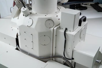
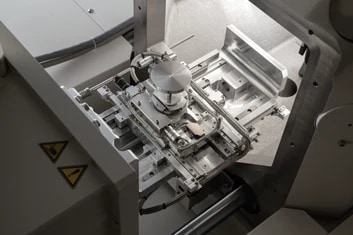
(*)at 0° tilt
Camera Navigation(*)
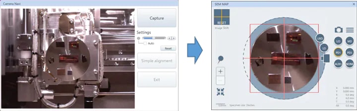
Right: Camera image transferred to the SEM MAP screen for navigation.
The camera navigation feature correlates an optical image to the target observation area.
The camera installed in the specimen chamber captures the specimen image at the time of specimen introduction. The image is transferred to the SEM MAP screen for a graphically driven navigation interface.
Camera navigation supports a maximum of φ 100 mm specimen.
(*)Camera navigation function is optional
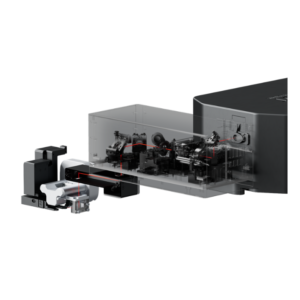
Detection System Enabling Dynamic Observation
The SU7000 supports observation under various environmental conditions. A variety of detectors (*) such as UVD and MD are selectable in addition to the PD-BSED for observation under low-vacuum conditions.
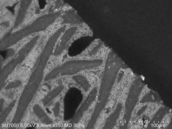
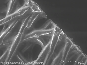
Detector Selection Under Low-Vacuum Conditions
Specimen: Fiber with metallic oxide
Left: MD (Backscattered electron) image
Right: UVD (SE image)
The oxide dispersion and fiber layering state are observed respectively.
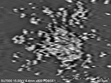
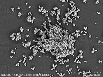
Improved PD-BSED Response Speed
Left: Traditional PD-BSED response at the scan rate of 30 ms x 64 frames
Right: SU7000 PD-BSED image demonstrating improved response and image quality to expand in-situ observation capability
Specifications
| Image Resolution | Resolution SE | 0.8 nm@15 kV | |
|---|---|---|---|
| 0.9 nm@1 kV | |||
| Magnification | 20~2,000,000 x | ||
| Electron Optics | Emitter | ZrO/W Schottky Emitter | |
| Accelerating Voltage | 0.1~30 kV (0.01 kV step) | ||
| Probe Current | Max. 200 nA | ||
| Detectors | Standard Detectors | UD(Upper Detector) | |
| MD(Middle Detector) | |||
| LD(Lower Detector) | |||
| Optional Detectors | PD-BSED(Semiconductor type) | ||
| UVD (Ultra Variable Pressure Detector) | |||
| Variable Pressure(VP) Mode (Option) | Pressure Range | 5~300 Pa | |
| Available Detectors in VP mode | PD-BSED, UVD, UD, MD,LD | ||
| Specimen Stage | Stage Control | 5-axis Motor Drive | |
| Movable Range | X | 0~135 mm | |
| Y | 0~100 mm | ||
| Z | 1.5~40 mm | ||
| T | -5~70° | ||
| R | 360° | ||
| Specimen Chamber | Specimen Size | Max. φ200 mm, Max. 67 mm Height | |
| Monitor(Option) | 23 inch LCD(1,920×1,080) , supports dual monitors operation | ||
| Image Display Mode | Large Screen Display Mode | 1,280×960 pixels | |
| Single Image Display Mode | 800×600 pixels | ||
| Dual Image Display Mode | 800×600 pixels、1,280×960 pixels with dual monitors | ||
| Quad Image Display Mode | 640×480 pixels | ||
| Hex Image Display Mode w/dual monitors | 640×480 pixels with dual monitors | ||
| Image Data Saving | Pixel Size | 640×480、1,280×960、2,560×1,920、5,120×3,840、10,240×7,680 | |
| Optional Accessories | Energy Dispersive X-ray Spectrometer (EDX) | ||
| Wavelength Dispersive X-ray Spectrometer (WDX) | |||
| Electron Backscattered Diffraction Detector (EBSD) | |||
| Cathodoluminescence System (CL) | |||
| Cryogenic Transfer System | |||
| Compatible with various types of sub-stages | |||
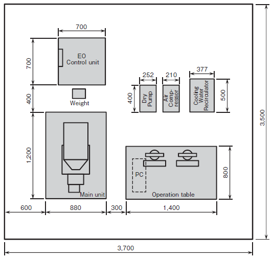
Installation Diagram (mm)


")
")