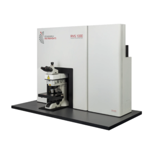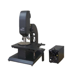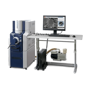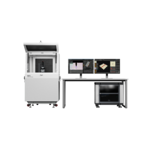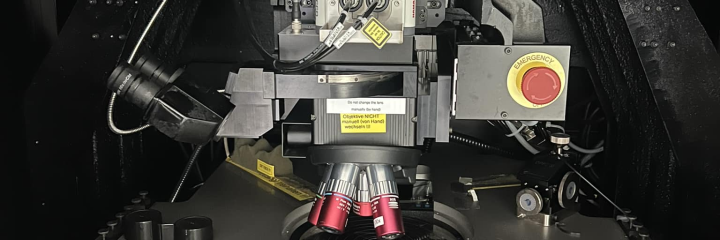

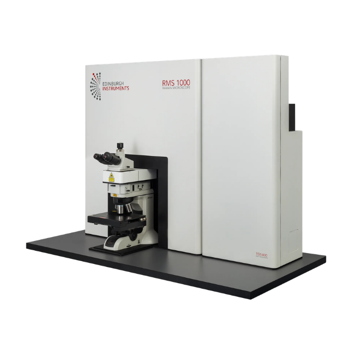
The RMS1000 is a Multimodal Confocal Raman Microscope built for adaptability and designed to grow, upgrade, and evolve with your research needs.
Built without compromise, this premium research tool offers unmatched specifications and ease of use. Its unique design allows full optimisation to any Raman microscopy application. With versatile laser, detector, and spectrograph options, the RMS1000 is perfect for any laboratory, finding applications in bioscience, nanomaterials, pharmaceuticals, semiconductors, geology, and more.
The RMS1000 is more than a Raman-only system. Harnessing Edinburgh Instruments’ extensive expertise in photoluminescence, we’ve integrated advanced fluorescence techniques to create a user-friendly, multimodal instrument. Seamlessly operate Raman and fluorescence lifetime imaging (FLIM) in a single software package, making RaFLIM® data acquisition from the same point both easy and intuitive.
Expanding on Raman and FLIM, the RMS1000 is exceptionally versatile, expandable to integrate a multitude of advanced techniques. Unlock precise imaging across various techniques with our multimodal system, featuring Raman, photoluminescence (PL), fluorescence life time imaging (FLIM), phosphorescence lifetime imaging (PLIM), multiphoton techniques, and antibunching capabilities.
The RMS1000 is a cutting-edge Multimodal Confocal Raman Microscope renowned for its adaptability and ease of upgrade. Its open architecture design facilitates seamless upgrades without compromising the high specifications of the core instrument. Built to adapt to your research needs, it integrates advanced techniques ensuring precise and comprehensive imaging capabilities across various scientific disciplines.
Raman Mapping
2D mapping and 3D mapping allow the user to see distribution of components, as well as revealing areas under high stress or strain, and defects. If using short exposure times FastMAP® is also available to reduce total acquisition times.
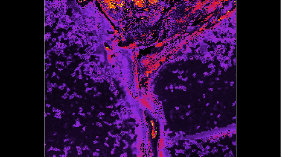
Figure 1: 2D Raman mapping showing nanocrystalline silicon and strain (pink, yellow, red) in a defective wafer
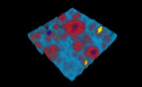
Figure 2: 3D Raman map of a pharmaceutical oil-in-water emulsion with TiO2 inclusions
SurfMAP®
SurfMAP® is a tool which allows accurate mapping of uneven surfaces giving the user assurance that their measurements are in focus and data is reliable. Ramacle builds a surface image which can be viewed in 2D and in 3D topography view. The software then acquires the spectral map moving the stage in X, Y, and Z for perfect laser focus.
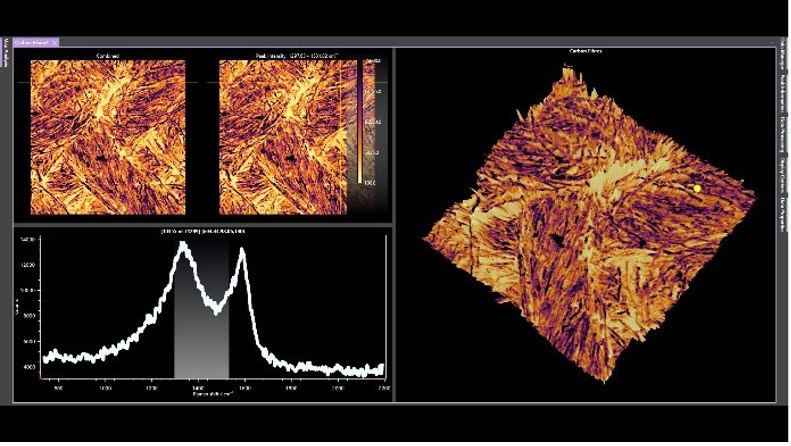
Figure 1: Raman SurfMAP® of carbon fibres tracking the D and G bands
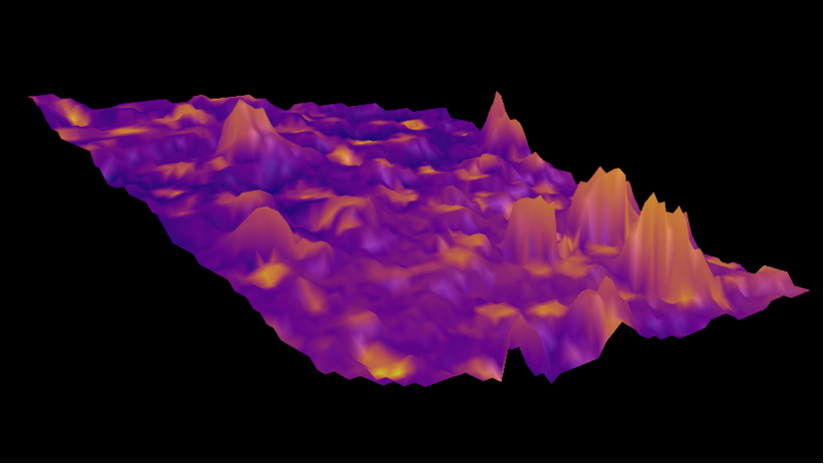
Figure 2: Raman SurfMap of an antipsychotic pharmaceutical drug
RaFLIM®
RaFLIM is a standout technique on the RMS1000. Combining Raman and FLIM to create maps of the same area, with the same spatial resolution (laser dependent). Operating measurement acquisition and data analysis in Ramacle for ease of use and continuity.
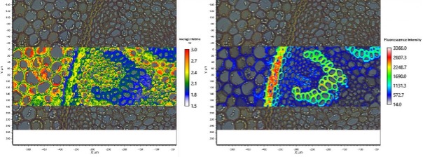
Figure 1: Average lifetime and fluorescence intensity image of a Convallaria Rhizome section stained with Acridine Orange. FLIM reveals variation in lifetime across lignified and pectin rich cell walls (2 exponential tail fitting).
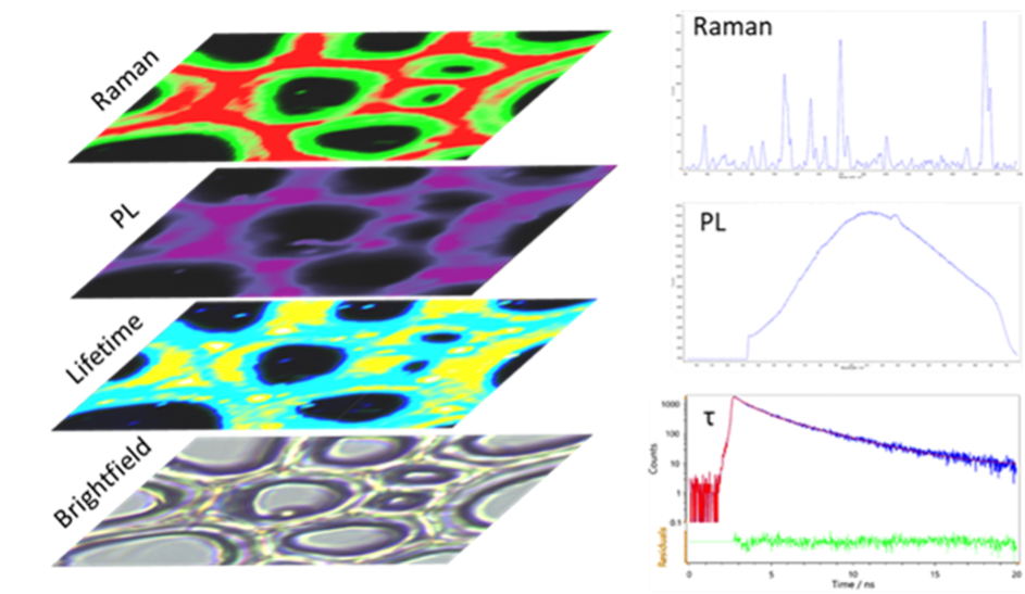
Figure 2: Biochemical analysis of woody plant cell walls using our multi-modal micro-spectroscopy approach.
Multimodal Techniques
The RMS1000’s external laser port unlocks limitless potential for advanced techniques like multiphoton imaging. Techniques such as two-photon excited fluorescence (2PEF) and second harmonic generation (SHG) are ideal for studying biological samples. Both 2PEF and SHG demand extremely high excitation intensity, provided by a mode-locked femtosecond pulsed laser.
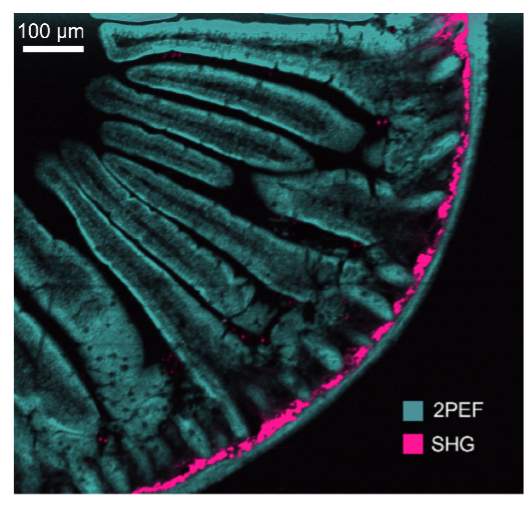
Figure 1: Mouse intestine stained with Alexa Fluor® 568
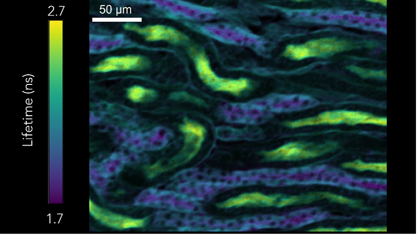
Figure 2: Photon lifetime imaging of mouse kidney sample
The sample of the mouse intestine (Figure 1) stained with Alexa Fluor® 568. Simultaneous measuring of spectral 2PEF and SHG from Alexa Fluor® 568 dye and fibrillar collagen respectively. Measurement using femtosecond laser and CCD camera. Using the same laser, two photon lifetime imaging was carried out an a sample of mouse kidney (Figure 2). This time a Hybrid Photodetector was used with TCSPC.
2D materials are also often studied with different measurement techniques, the RMS1000 can easily be configured to carry out all imaging techniques on the same sample area providing a full suite of complimentary information. Raman and PL images were collected using a 532 nm laser, whilst the SHG was acquired using a femtosecond laser.
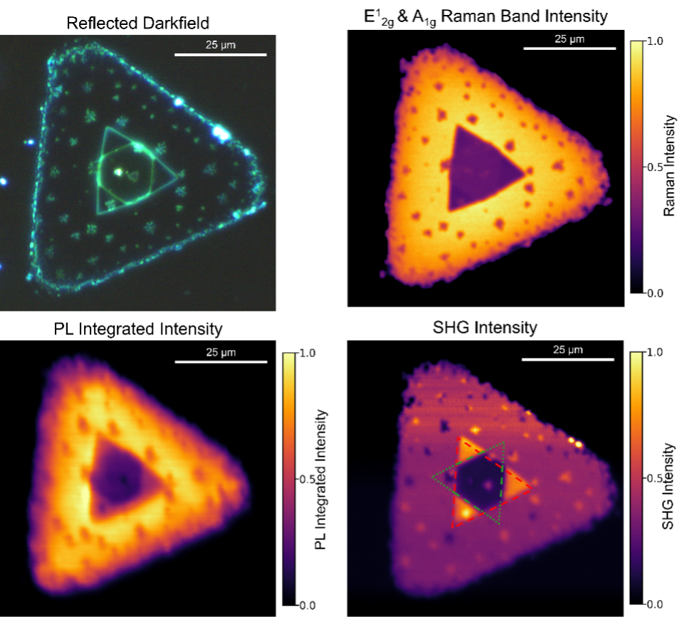
Figure 3: Sample of WSe2 crystal. The darkfield (DF) image reveals areas of different heights. Raman, PL, and SHG images can be used correlatively to identify layer number, and highlight areas of stress, strain, dopants, and defects